Rib Cage Muscles Anatomy : Ribs Pictures Anatomy Anatomy Body Maps : Rib 2 is thinner and longer than rib 1 and has two articular facets on the head as normal.
Rib Cage Muscles Anatomy : Ribs Pictures Anatomy Anatomy Body Maps : Rib 2 is thinner and longer than rib 1 and has two articular facets on the head as normal.. 1887 human anatomy print of the rib cage and sternum. It has clear front side and back planes. Sign up for premium today! Volume rendering of a contrast enhanced thoracoabdominal ct scan. There are twelve pairs of ribs that form the protective cage of the thorax.
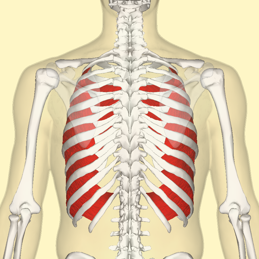
Serratus posterior superior and inferior. Each rib articulates posteriorly with the vertebral column. The rib cage surrounds the lungs and the heart, serving as an important means of bony protection for these vital organs. Your rib cage plays an important role in respiration, expanding and contracting as your respiratory muscles, including your diaphragm, work to help you breathe. The rib cage is composed by sternum, costal cartilages, and ribs connected to the thoracic vertebrae.
Internal Intercostal Muscles Wikipedia from upload.wikimedia.org For a gesture drawing, that's good enough. Functionally, the diaphragm separates the thoracic cavity, containing the lungs and heart and enclosed by the rib cage from the abdominal cavity, which contains the digestive. On a muscular person when the muscles stretch, we see some of the lower ribs in the front and also in the back. Structure and function (6th ed.). The rib cage is a primarily protective structure, encircling the heart and lungs. The ribs joint as follows muscles of the thoracic wall contain those that fill and support the intercostal spaces, those that pass between the sternum and the ribs, and those that cross several. In this lesson i review and critique your assignments on the rib cage. Muscle spasms located in the rib cage are often observed in people who strain or overwork their upper body muscles.
Rib cage anatomy and breathing.
Ribs & thoracic cage muscles attachments. Anatomy of the human body for artists. Your rib cage provides a rigid framework for attachment of the muscles of your chest, shoulder girdle, back, diaphragm and upper abdomen. The intercostal muscles are the muscles that occupy the 11 intercostal spaces. They are more involved in forced expiration and coughing to forcibly shrink the chest and. Structure of a typical rib: • raise rib cage for inhaling & depresses rib cage for exhaling. The thorax is anatomical structure supported by a skeletal framework (thoracic cage) and the ribs on both the sides complete the cage. Rib cage anatomy and breathing. For a gesture drawing, that's good enough. The thoracic cage (rib cage) is the skeletal framework of the thoracic wall, which encloses the thoracic cavity. The rib cage is often simplified as an oval shape. Ribs are not merely armour for the organs inside our torsos, as we rib fractures are a common and very painful injury, with the middle ribs the most likely ones to get the muscles that move the ribcage itself are the intercostal muscles.
• raise rib cage for inhaling & depresses rib cage for exhaling. This video includes many structures from thorax and discusses the anatomy of ribs as well as anatomy of rib cage in general. The rib cage, shaped in a mild cone shape and more flexible than most bone sets, is made up of varying elements such as the thoracic vertebra, 12 equally paired ribs, costal cartilage, and held together anteriorly by the sternum. Structure of a typical rib: See more ideas about anatomy, anatomy study, rib cage anatomy.
Rib Cage Muscles Images Stock Photos Vectors Shutterstock from image.shutterstock.com There are twelve pairs of ribs that form the protective cage of the thorax. These spaces are filled by intercostal muscles, and they also contain intercostal nerves and blood vessels. Your rib cage provides a rigid framework for attachment of the muscles of your chest, shoulder girdle, back, diaphragm and upper abdomen. Everyone has nice muscles in ct scanning! Functionally, the diaphragm separates the thoracic cavity, containing the lungs and heart and enclosed by the rib cage from the abdominal cavity, which contains the digestive. These muscles may be located anteriorly, posteriorly, and/or laterally. Anterior view of muscle attachments of chest costa. Muscles that move the rib cage attach to the rib cage.
Structure and function (6th ed.).
Abdomen & ribs muscle movements. Some extend from above and draw the. So what parts of the rib cage show up on the surface? Ribs & thoracic cage muscles attachments. Illustration of thoracic vertebrae showing vertebral body, pedicles, facets, transverse process, rib. For a gesture drawing, that's good enough. Rib cage anatomy and breathing. The rib cage is a primarily protective structure, encircling the heart and lungs. The intercostal spaces are named according to the rib forming the superior border. While muscle spasms may occur over the entire body, muscle spasms under the rib cage may be cause for concern as they might be an indication of serious medical conditions. 1887 human anatomy print of the rib cage and sternum. Learn about ribs muscle with free interactive flashcards. Structure and function (6th ed.).
Sign up for premium today! Volume rendering of a contrast enhanced thoracoabdominal ct scan. Rib 2 is thinner and longer than rib 1 and has two articular facets on the head as normal. Your rib cage plays an important role in respiration, expanding and contracting as your respiratory muscles, including your diaphragm, work to help you breathe. Structure and function (6th ed.).
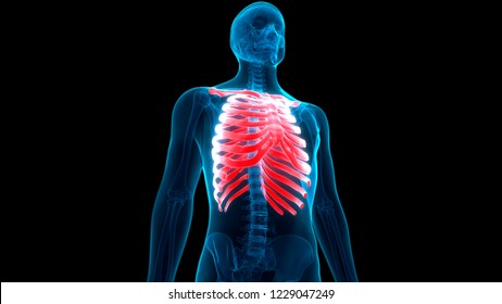
Intercostal Muscles Anatomy Stock Illustration 42176740 Pixta from en.pimg.jp During normal breathing, contraction of the major inspiratory muscle, the diaphragm, produces both rib cage expansion and a downward movement of the diaphragm. Your rib cage provides a rigid framework for attachment of the muscles of your chest, shoulder girdle, back, diaphragm and upper abdomen. Rib cage anatomy and breathing. Anatomy of the human body for artists. Sign up for premium today! Illustration of thoracic vertebrae showing vertebral body, pedicles, facets, transverse process, rib. They are more involved in forced expiration and coughing to forcibly shrink the chest and. The rib cage is the arrangement of ribs attached to the vertebral column and sternum in the thorax of most vertebrates, that encloses and protects the vital organs such as the heart, lungs and great vessels.
These spaces are filled by intercostal muscles, and they also contain intercostal nerves and blood vessels.
The rib cage is often simplified as an oval shape. Various skeletal muscles are attached to the rib cage. Learn about ribs muscle with free interactive flashcards. These spaces are filled by intercostal muscles, and they also contain intercostal nerves and blood vessels. Each rib articulates posteriorly with the vertebral column. The ribs joint as follows muscles of the thoracic wall contain those that fill and support the intercostal spaces, those that pass between the sternum and the ribs, and those that cross several. The rib cage surrounds the lungs and the heart, serving as an important means of bony protection for these vital organs. Sign up for premium today! Skeletal muscles attached to the rib cage: This video includes many structures from thorax and discusses the anatomy of ribs as well as anatomy of rib cage in general. This cage protects vital organs and is essential for creating negative pressure to inflate lungs. For a gesture drawing, that's good enough. The other attachment of these muscles is usually considered to be either superior or inferior to the rib attachment.


0 Comments